A Multimodal Dataset with EEG and forehead EOG for Resting-State analysis.
This dataset is a subset of SPIS Resting-State EEG Dataset.
Copyright:
Mastaneh Torkamani-Azar and Mujdat Cetin
Signal Processing and Information Systems (SPIS) Laboratory
Faculty of Engineering and Natural Sciences
Sabanci University
Istanbul 34956, Turkey
If you use SPIS Resting-State Dataset in your research, please cite the following paper:
M. Torkamani-Azar, S. D. Kanik, S. Aydin and M. Cetin, "Prediction of Reaction Time and Vigilance Variability From Spatio-Spectral Features of Resting-State EEG in a Long Sustained Attention Task," in IEEE Journal of Biomedical and Health Informatics, vol. 24, no. 9, pp. 2550-2558, Sept. 2020, doi: 10.1109/JBHI.2020.2980056. https://ieeexplore.ieee.org/document/9034192
If you have any questions about this dataset, please kindly contact Dr. Mastaneh Torkamani Azar (mastaneh.torkamani@uef.fi, She/Her) or Prof. Mujdat Cetin (mujdat.cetin@rochester.edu, He/Him). http://labs.sabanciuniv.edu/spis/
These 10 datasets were recorded prior to a 105-minute session of Sustained Attention to Response Task with fixed-sequence and varying ISIs.
Each dataset contains 2.5 minutes of eyes-open (EO) and 2.5 minutes of eyes-closed (EC) resting-state EEG.
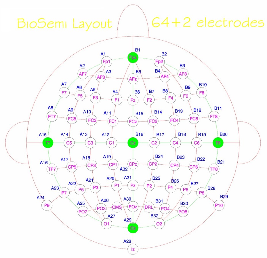
Monopolar EEG activity was collected at 2,048 Hz via 64 Ag/AgCl active electrodes mounted according to the 10-10 International Electrode Placement System.
Experiments were conducted in the early afternoon to induce drowsiness in the already idled brain networks.
All files are saved as the .mat format using Matlab Version 7.3. The channe location is also provided.
The raw data in this file has been downsampled to 256 Hz from the original sampling rate of 2,048 Hz.
Channels 1 to 64 correspond to the monopolar EEG channels using the following channel names:
channelList = {'Fp1';'AF7';'AF3';'F1';'F3';'F5';'F7';'FT7';'FC5';'FC3';'FC1';'C1';'C3';'C5';'T7';'TP7';'CP5';'CP3';'CP1';'P1';'P3';'P5';'P7';. .. 'P9';'PO7';'PO3';'O1';'Iz';'Oz';'POz';'Pz';'CPz';'Fpz'; 'Fp2';'AF8';'AF4';'Afz';'Fz';'F2';'F4';'F6';'F8';'FT8';'FC6';'FC4';'FC2';'FCz';'Cz';... 'C2';'C4';'C6';'T8';'TP8';'CP6';'CP4';'CP2';'P2';'P4';'P6';'P8';'P10';'PO8';'PO4';'O2'};
Channels 65 to 67: Three surface EOG electrodes connected to the outer corners of the eyes and middle of the eyebrows.
Channel 68: The trigge channel.
trigger values = 200 correspond to the eyes-open (EO) and trigger values = 220 correspond to the eyes-closed (EC) states.
The data was collected using a 64-channel Biosemi Active 2 amplifier and the resulting raw signals are measured in microvolts. You can also adjust the channels for their gain if it is important for your calculations. A very good reference on Biosemi channel characteristics can be found at https://www.fieldtriptoolbox.org/getting_started/biosemi/. In addition, quoting from https://www.biosemi.com/faq/cms&drl.htm, "The signals on file are the voltages between each electrode and CMS". In other words, raw voltages are not exactly measured with respect to a ground or reference electrode but with respect to a common mode sense electrode. Both CMS and DRL are located on the parietooccipital area. You can check the layout from this link: https://www.biosemi.com/pics/cap_64_layout_medium.jpg.
Please refer to the methods in the source paper or articles that have developed artifact removal technique using this dataset: https://scholar.google.com/scholar?cites=2250172710784316618&as_sdt=2005&sciodt=0,5&hl=en